Scanning electron microscope images of beaded Sepharose CL-6B with 100
Par un écrivain mystérieux
Description
Confocal microscopy analysis reveals that only a small proportion of extracellular vesicles are successfully labelled with commonly utilised staining methods

Enhancing Extracellular Vesicle Analysis by Integration of Large-Volume Sample Stacking in Capillary Electrophoresis with Asymmetrical Flow Field-Flow Fractionation

Affinity-Based Magnetic Particles for the Purification of Single-Stranded DNA Scaffolds for Biomanufacturing DNA-Origami Nanostructures

Biotechnology & Bioengineering, Biotechnology Journal
Detection and phenotyping of extracellular vesicles by size exclusion chromatography coupled with on-line fluorescence detection
Biology, Free Full-Text
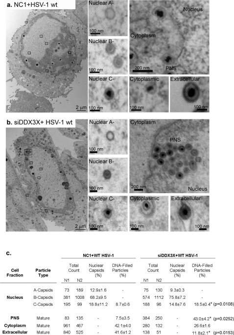
RNA helicase DDX3X modulates herpes simplex virus 1 nuclear egress

One-Step Fabrication of Hollow Spherical Cellulose Beads: Application in pH-Responsive Therapeutic Delivery
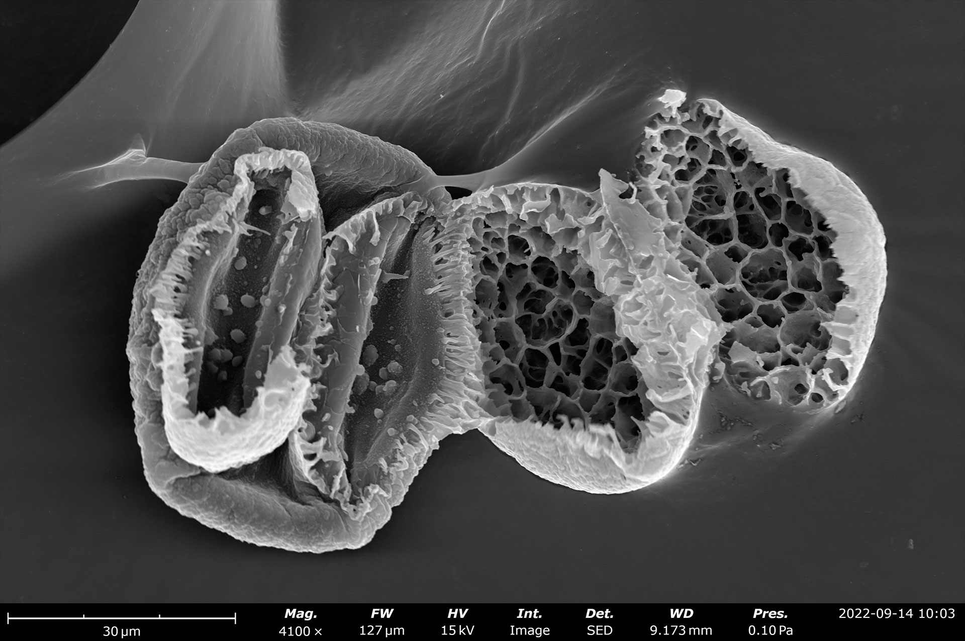
Scanning Electron Microscopy
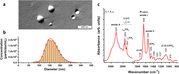
Improved isolation of extracellular vesicles by removal of both free proteins and lipoproteins
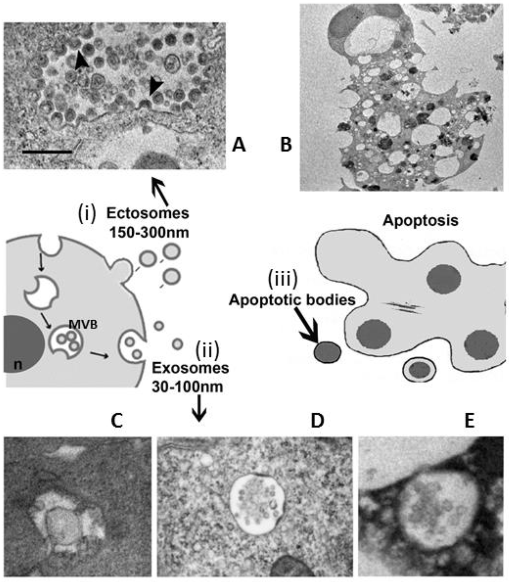
Frontiers MicroRNAs in extracellular vesicles: Sorting mechanisms, diagnostic value, isolation, and detection technology

Representative SEM micrographs of Sepharose beads at 150 × (A), 450 ×

αS1-Casein-Loaded Proteo-liposomes as Potential Inhibitors in Amyloid Fibrillogenesis: In Vivo Effects on a C. elegans Model of Alzheimer's Disease

Scanning electron microscope images of beaded Sepharose CL-6B with 100
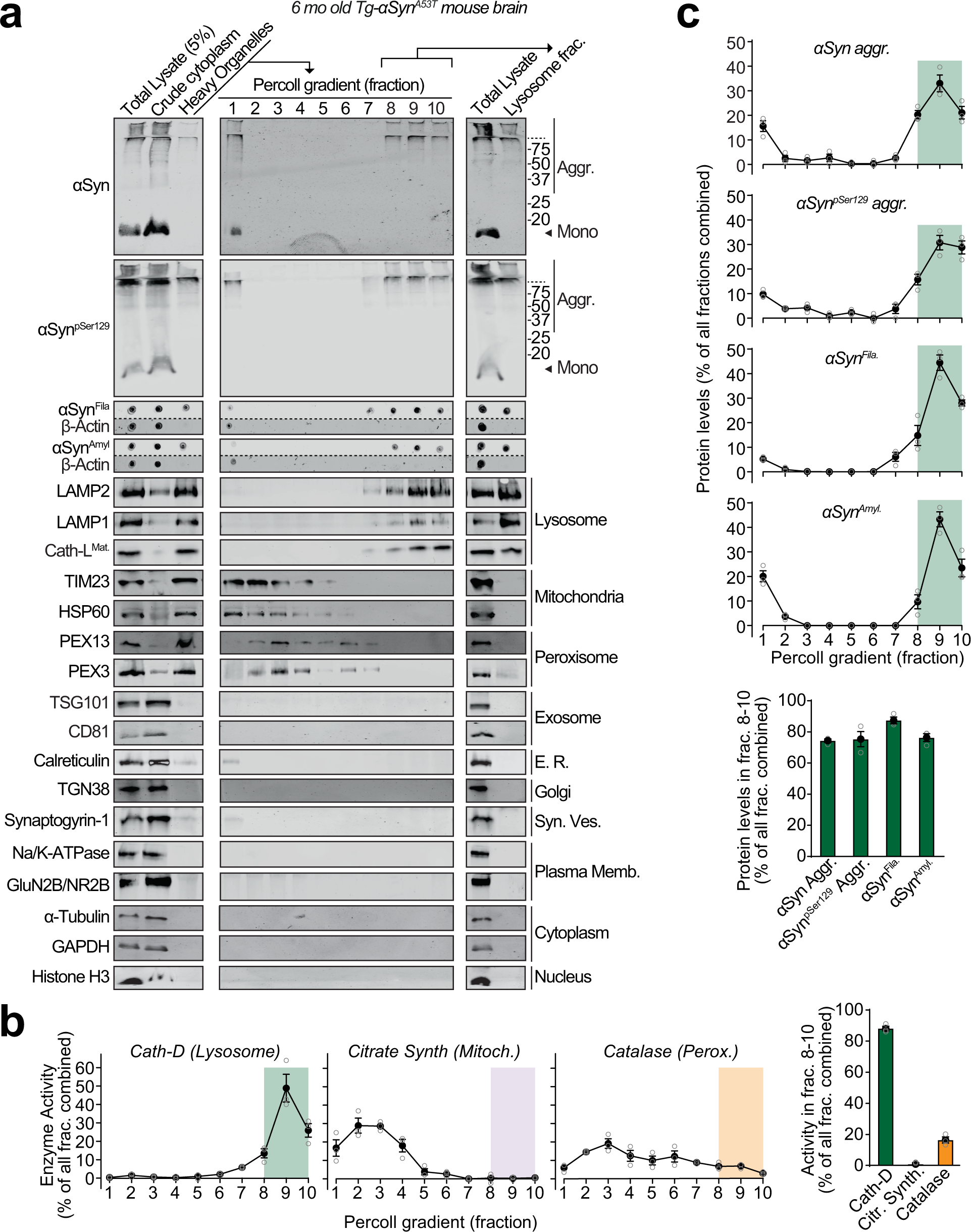
Lysosomal exocytosis releases pathogenic α-synuclein species from neurons in synucleinopathy models
depuis
par adulte (le prix varie selon la taille du groupe)